International Journal of Computer Assisted Radiology and Surgery
Cardiac MRI visualization for ventricular tachycardia ablation
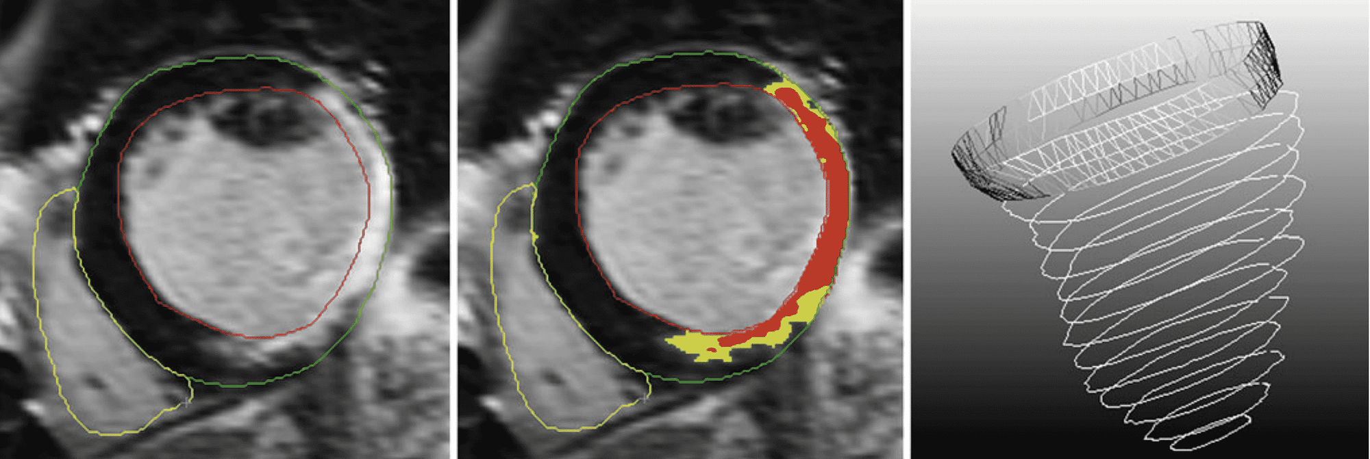
Objective The integrated visualization of cardiac MRI during a ventricular tachycardia (VT) mapping and ablation procedure would provide improved catheter guidance and tissue assessment. We developed a system for and explored the added value of simultaneous visualization of intracardiac voltage measurements and MRI-derived myocardial scar information during VT ablation procedures. Method We propose the use of a synchronized 3D and 2D view. In 3D, the catheter will be guided optimally by assessing 3D scar characteristics and its relation to the ventricular anatomy. In 2D, a detailed assessment of the tissue can be made. We developed several 3D visualization techniques, including volume rendering of the scar and myocardial surfaces colored according to the voltage measurements. We also visualized context structures in the heart. For the 2D view, we proposed showing three adjacent slices simultaneously. To link the 3D with the 2D view, we added a linking plane and linking contours; the slice level shown in the 2D view is indicated in the 3D view. Results We evaluated our method via a case study during which we simulated the visual environment of an ablation procedure. The MRI-based volume rendering of scar tissue and the linking between the 3D and 2D views were both positively received. However, the visualization of the voltage measurements was found to be hard to interpret, partly due to the perceptually suitable but non-standard colormap. Conclusions Based on this study, we can conclude that our approach of displaying MRI data and integrating it with voltage measurements has potential to improve VT ablation procedures.
More Information
Citation
BibTex
@article{bib:godeschalk-slagboom:2012,
    author       = { Godeschalk-Slagboom, Corine and van der Geest, Rob and Zeppenfeld, Katja and Botha, Charl P. },
    title        = { Cardiac MRI visualization for ventricular tachycardia ablation },
    journal      = { International Journal of Computer Assisted Radiology and Surgery },
    volume       = { 7 },
    year         = { 2012 },
    pages        = { 753--767 },
    doi          = { 10.1007/s11548-012-0776-4 },
    dblp         = { journals/cars/Godeschalk-SlagboomGZB12 },
    url          = { https://publications.graphics.tudelft.nl/papers/285 },
}