Skeletal Radiol
Measuring femoral lesions despite CT metal artefacts: a cadaveric study
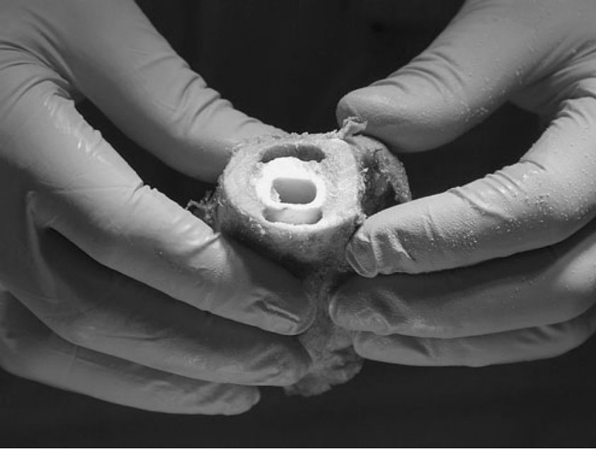
Objective Computed tomography is the modality of choice for measuring osteolysis but suffers from metal-induced artefacts obscuring periprosthetic tissues. Previous papers on metal artefact reduction (MAR) show qualitative improvements, but their algorithms have not found acceptance for clinical applications. We investigated to what extent metal artefacts interfere with the segmentation of lesions adjacent to a metal femoral implant and whether metal artefact reduction improves the manual segmentation of such lesions. Materials and methods We manually created 27 periprosthetic lesions in 10 human cadaver femora. We filled the lesions with a fibrotic interface tissue substitute. Each femur was fitted with a polished tapered cobalt-chrome prosthesis and imaged twice — once with the metal, and once with a substitute resin prosthesis inserted. Metal-affected CTs were processed using standard back-projection as well as projection interpolation (PI) MAR. Two experienced users segmented all lesions and compared segmentation accuracy. Results. We achieved accurate delineation of periprosthetic lesions in the metal-free images. The presence of a metal implant led us to underestimate lesion volume and introduced geometrical errors in segmentation boundaries.
More Information
Citation
BibTex
@article{bib:malan:2012,
    author       = { Malan, Daniel F. and Botha, Charl P. and Kraaij, Gert and Joemai, Raoul M. S. and van der Heide, Huub J. L. and Nelissen, Rob. G. H. H. and Valstar, Edward R. },
    title        = { Measuring femoral lesions despite CT metal artefacts: a cadaveric study },
    journal      = { Skeletal Radiol },
    volume       = { 41 },
    year         = { 2012 },
    pages        = { 547--55 },
    doi          = { 10.1007/s00256-011-1223-2 },
    pubmedid     = { 21732221 },
    url          = { https://publications.graphics.tudelft.nl/papers/396 },
}