Catheter Cardiovasc Interv
Fully automatic three-dimensional quantitative analysis of intracoronary optical coherence tomography: method and Validation
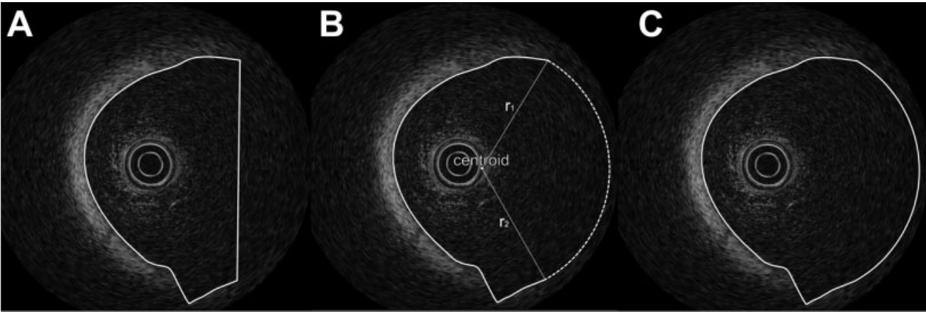
Quantitative analysis of intracoronary optical coherence tomography (OCT) image data (QOCT) is currently performed by a time-consuming manual contour tracing process in individual OCT images acquired during a pullback procedure (frame-based method). To get an efficient quantitative analysis process, we developed a fully automatic three-dimensional (3D) lumen contour detection method and evaluated the results against those derived by expert human observers. Methods: The method was developed using Matlab (The Mathworks, Natick, MA). It incorporates a graphical user interface for contour display and, in the selected cases where this might be necessary, editing. OCT image data of 20 randomly selected patients, acquired with a commercially available system (Lightlab imaging, Westford, MA), were pulled from our OCT database for validation. Results: A total of 4,137 OCT images were analyzed. There was no statistically significant difference in mean lumen areas between the two methods (5.03 +- 2.16 vs. 5.02 +- 2.21 mm power 2; P = 0.6, human vs. automated). Regression analysis showed a good correlation with an r value of 0.99. The method requires an average 2–5 sec calculation time per OCT image. In 3% of the detected contours an observer correction was necessary. Conclusion: Fully automatic lumen contour detection in OCT images is feasible with only a select few contours showing an artifact (3%) that can be easily corrected. This QOCT method may be a valuable tool for future coronary imaging studies incorporating OCT.
More Information
Citation
BibTex
@article{bib:sihan:2009,
    author       = { Sihan, K. and Botha, Charl P. and Post, Frits H. and de Winter, Sebastiaan and Nieves, Gonzalo and Regar, E and Serruys, Patrick J W C and Hamers, R and Bruining, N },
    title        = { Fully automatic three-dimensional quantitative analysis of intracoronary optical coherence tomography: method and Validation },
    journal      = { Catheter Cardiovasc Interv },
    volume       = { 74 },
    year         = { 2009 },
    pages        = { 1058--65 },
    doi          = { 10.1002/ccd.22125 },
    pubmedid     = { 19521990 },
    url          = { https://publications.graphics.tudelft.nl/papers/421 },
}