International Journal of Computer Assisted Radiology and Surgery
Voxel classification and graph cuts for automated segmentation of pathological periprosthetic hip anatomy
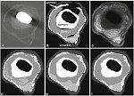
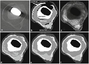
Purpose Automated patient-specific image-based segmentation of tissues surrounding aseptically loose hip prostheses is desired. For this we present an automated segmentation pipeline that labels periprosthetic tissues in computed tomography (CT). The intended application of this pipeline is in pre-operative planning. Methods Individual voxels were classified based on a set of automatically extracted image features. Minimum-cost graph cuts were computed on the classification results. The graph-cut step enabled us to enforce geometrical containment constraints, such as cortical bone sheathing the femur’s interior. The solution’s novelty lies in the combination of voxel classification with multilabel graph cuts and in the way label costs were defined to enforce containment constraints. Results The segmentation pipeline was tested on a set of twelve manually segmented clinical CT volumes. The distribution of healthy tissue and bone cement was automatically determined with sensitivities greater than 82% and pathological fibrous interface tissue with a sensitivity exceeding 73%. Specificity exceeded 96% for all tissues. Conclusions The addition of a graph-cut step improved segmentation compared to voxel classification alone. The pipeline described in this paper represents a practical approach to segmenting multitissue regions from CT.
More Information
Gallery
Citation
BibTex
@article{bib:malan:2013,
    author       = { Malan, Daniel F. and Botha, Charl P. and Valstar, Edward R. },
    title        = { Voxel classification and graph cuts for automated segmentation of pathological periprosthetic hip anatomy },
    journal      = { International Journal of Computer Assisted Radiology and Surgery },
    volume       = { 8 },
    year         = { 2013 },
    pages        = { 63--74 },
    doi          = { 10.1007/s11548-012-0671-z },
    dblp         = { journals/cars/MalanBV13 },
    url          = { https://publications.graphics.tudelft.nl/papers/564 },
}